
-
Chemiluminescent Western blotting
- AdvanStain Iris
- WesternEaze-Chemi Kit
- AdvanStain Total Fluorescent Protein Staining Kits
- AdvanBlock-Chemi blocking solution
- FLASHBlot transfer buffer
- FLASHBlot-SD transfer buffer
- Development folders
- WesternBright ECL HRP substrate
- WesternBright ECL Spray
- WesternBright Quantum HRP substrate
- WesternBright Sirius HRP substrate
- HRP-conjugated secondary antibodies
- LucentBlue X-ray film
- LucentBlue X-ray film (CLONE)
- LucentBlue X-ray film (CLONE)
- Background Quenching Sheets
- WesternBright ChemiPen
- Transfer membranes
- Incubation trays
- AdvanWash washing solution
- Western Blot Strip-It Buffer
- Blotting sponge pads
- Blotting papers
- X-ray film cassette
-
Fluorescent Western blotting
- FLASHBlot transfer buffer
- AdvanBlock-Fluor blocking solution
- AdvanBlock-PF blocking solution
- AdvanStain Total Fluorescent Protein Staining Kits
- LightSaver™ Fluorescence Enhancing Solution
- FLASHBlot-SD transfer buffer
- AdvanStain Iris
- WesternBright MCF
- SpectraDye Secondary Antibodies
- SpectraDye Antibody Labeling Kits
- Background Quenching Sheets
- Transfer membranes
- Development folders
- Incubation trays
- AdvanWash washing solution
- Fluorescent Western standardization blot
- Blotting papers
- ELISA
- Electrophoresis
- Antibodies and antibody labeling
- Sample preparation
- Purification
- Protein staining
-
Buffers and solutions
- FLASHBlot transfer buffer
- AdvanBlock-PF blocking solution
- Western Blot Strip-It Buffer
- AdvanBlock-Chemi blocking solution
- AdvanBlock-EIA blocking solution
- AdvanBlock-Fluor blocking solution
- Protein sample loading buffers
- FLASHBlot-SD transfer buffer
- Cleanera Endotoxin Removal Buffer Kit
- AdvanWash washing solution
- 10X EIA Coating Buffer
- LightSaver™ Fluorescence Enhancing Solution
- Avant buffer pouches
- SARS-CoV-2
- Cell biology
- Lab supplies
- Custom services
New special offers are available!
AdvanStain Iris
Upgrade your protein staining! Boost sensitivity,
no-destaining, and high-contrast.
- Detection limit – 2 ng vs Ponceau at 150 ng
- Quantitative – effective for lower protein concentrations
- No destaining – allows direct chemiluminescent or fluorescent Western blotting without the need for destaining
- Versatile – compatible with both nitrocellulose and PVDF membranes


| CAT # | PRODUCT | SIZE | PRICE | QUANTITY | |
|---|---|---|---|---|---|
| R-03732-D25 | AdvanStain Iris | 250 ml |
$ 171.00 (USD) |
Description
Ponceau staining can be a headache – background signal often overlaps with your bands of interest, and when you try to reduce non-specific signal, your bands fade away. Achieving the rigth balance feels like a guessing game, especially with low protein concentrations. Enter AdvanStainTM IrisTM – your solution for frustration-free staining. With AdvanStain Iris, just stain, rinse, and visualize. No need for destaining or worrying about signal loss, saving you valuable time and effort. Its deep blue color provides exceptional contrast, ensuring crystal-clear protein band detection every time.
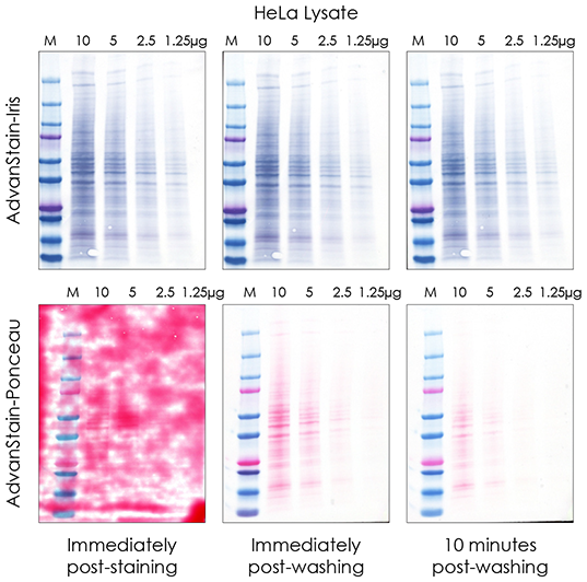
Flexible imaging, no destaining required

Figure 1. AdvanStain Iris demonstrates high sensitivity staining of HeLa lysate, no destaining is required prior to analysis by Western blot. AdvanStain Iris stain was applied to a nitrocellulose membrane for 10 minutes. (a) EPI Blue image (b) Visible image. Protein bands were observed in the 80ng lysate load. (c) After Staining, the membrane was blocked then probed with mouse anti-Vinculin (Boster #MA1103) followed by Goat anti-Mouse HRP (Advansta #R-05071-500). The Western blot was developed with WesternBright™ ECL Substrate (Advansta #K-12045).
Maximum sensitivity
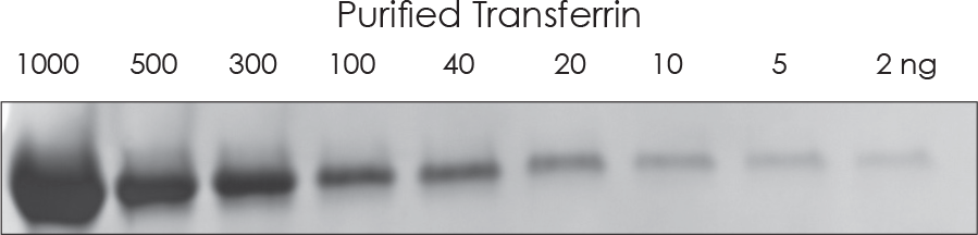
Figure 2. High sensitivity stain detects as little as 2 ng of purified protein per band. Dilutions of purified transferrin protein were electrophoresed using SDS-PAGE and the protein was transferred to a nitrocellulose membrane then stained with AdvanStain Iris for 10 minutes. Image was acquired with EPI Blue light.
Saves time and outperforms Ponceau S
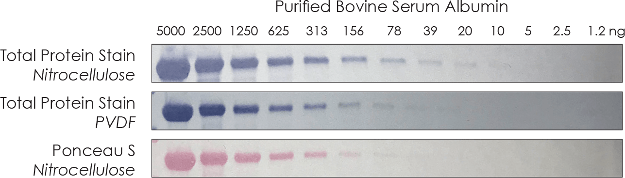
Figure 3. AdvanStain Iris is significantly more sensitive when compared to Ponceau S and compatible with PVDF membranes.Dilutions of purified BSA protein were electrophoresed using SDS-PAGE and the protein was transferred to a nitrocellulose or PVDF membrane then stained with AdvanStain Iris for 10 minutes or Ponceau S for 5 minutes after a 10 minute water rinse. Images were acquired with visible light.
Re-use up to 3X
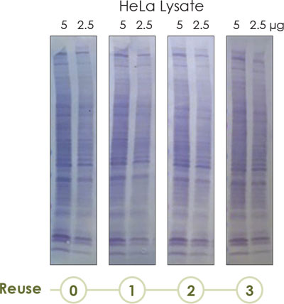
Figure 4. AdvanStain Iris may be re-used three times.Dilutions of HeLa Lysate were electrophoresed using SDS-PAGE and the protein was transferred to a nitrocellulose membrane then stained with AdvanStain Iris for 10 minutes. The staining solution was re-used three times to generate comparable data. Images were acquired with visible light.
Connect