-
Chemiluminescent Western blotting
- AdvanStain Iris
- WesternEaze-Chemi Kit
- AdvanStain Total Fluorescent Protein Staining Kits
- AdvanBlock-Chemi blocking solution
- FLASHBlot transfer buffer
- FLASHBlot-SD transfer buffer
- Development folders
- WesternBright ECL HRP substrate
- WesternBright ECL Spray
- WesternBright Quantum HRP substrate
- WesternBright Sirius HRP substrate
- HRP-conjugated secondary antibodies
- LucentBlue X-ray film
- Background Quenching Sheets
- WesternBright ChemiPen
- Transfer membranes
- Incubation trays
- AdvanWash washing solution
- Western Blot Strip-It Buffer
- Blotting sponge pads
- Blotting papers
- X-ray film cassette
-
Fluorescent Western blotting
- FLASHBlot transfer buffer
- AdvanBlock-Fluor blocking solution
- AdvanBlock-PF blocking solution
- AdvanStain Total Fluorescent Protein Staining Kits
- LightSaver™ Fluorescence Enhancing Solution
- FLASHBlot-SD transfer buffer
- AdvanStain Iris
- WesternBright MCF
- SpectraDye Secondary Antibodies
- SpectraDye Antibody Labeling Kits
- Background Quenching Sheets
- Transfer membranes
- Development folders
- Incubation trays
- AdvanWash washing solution
- Fluorescent Western standardization blot
- Blotting papers
- ELISA
- Electrophoresis
- Antibodies and antibody labeling
- Sample preparation
- Purification
- Protein staining
-
Buffers and solutions
- FLASHBlot transfer buffer
- AdvanBlock-PF blocking solution
- Western Blot Strip-It Buffer
- AdvanBlock-Chemi blocking solution
- AdvanBlock-EIA blocking solution
- AdvanBlock-Fluor blocking solution
- Protein sample loading buffers
- FLASHBlot-SD transfer buffer
- Cleanera Endotoxin Removal Buffer Kit
- AdvanWash washing solution
- 10X EIA Coating Buffer
- LightSaver™ Fluorescence Enhancing Solution
- Avant buffer pouches
- SARS-CoV-2
- Cell biology
- Custom services
AdvanStain Total Fluorescent Protein Staining Kits
Rapid fluorescent stain for total protein normalization
- Convenient – simple protocol that takes less than 20 minutes, no de-staining required
- Versatile – compatible with chemiluminescent and IR700/IR800 fluorescent Western blotting
- Accurate – linear across a broad range from 1-50μg of lysate, ideal for total protein normalization
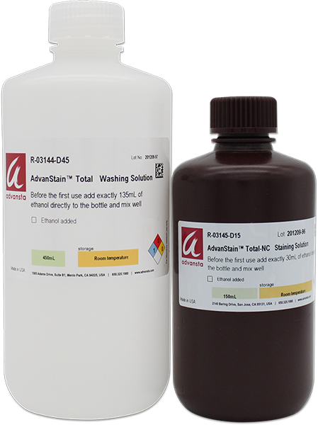
| CAT # | PRODUCT | SIZE | PRICE | QUANTITY | |
|---|---|---|---|---|---|
| K-11061-015 | AdvanStain Total-PVDF Fluorescent Protein Staining Kit | 15 blots |
$ 190.00 (USD) |
||
| K-11062-015 | AdvanStain Total-NC Fluorescent Protein Staining Kit | 15 blots |
$ 190.00 (USD) |
Description
AdvanStain Total is a fluorescent protein stain for PVDF and Nitrocellulose membranes that allows for sensitive and quantitative visualization of protein bands.
For precise quantitative Western blot data, it is critical to perform normalization to account for sample inconsistencies, sample loading errors and uneven transfer. In an effort to increase transparency, total protein normalization is quickly becoming the standard for Western blot normalization as it is now the preferred method for top tier journals. The AdvanStain Total Fluorescent Protein Staining Kits provide a convenient method for total protein normalization that is compatible with digital imaging systems equipped with a Cy3‑compatible filter.
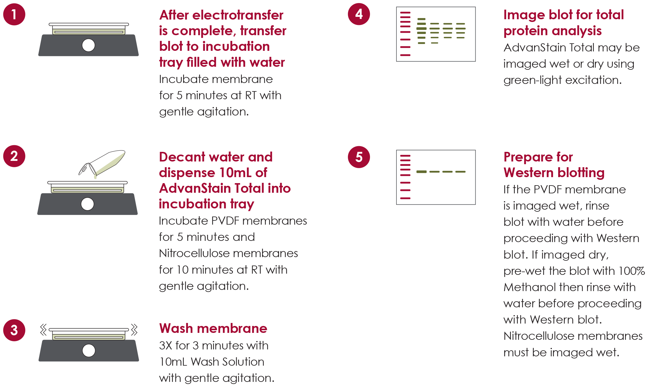
AdvanStain Total Fluorescent Protein Staining Workflow
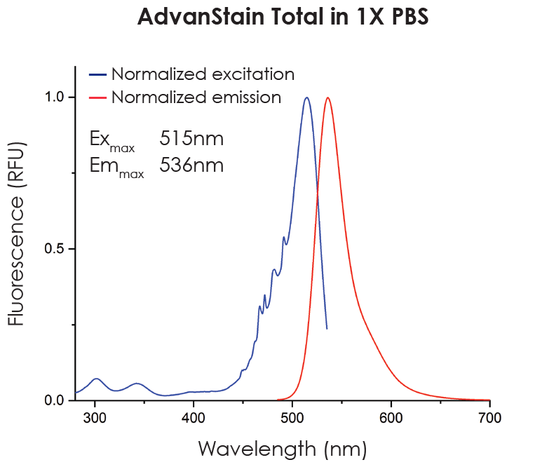
AdvanStain Total spectrum
Figure 1. AdvanStain Total can be imaged with a CCD or laser based imaging system using green-light excitation.
Broad linear range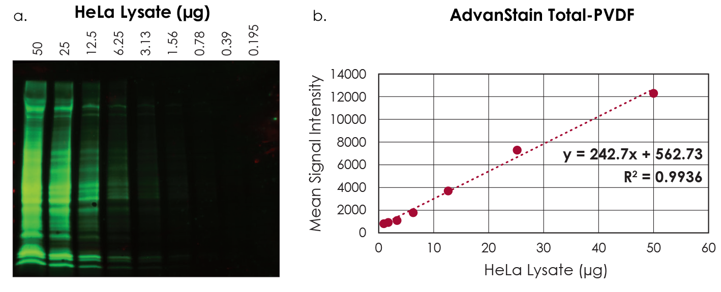
Figure 2. AdvanStain Total has a broad linear range. Untreated Hela lysate was serially diluted 1:2 prior to separation by SDS-PAGE. After electrophoresis the proteins were transferred to a PVDF membrane for total protein staining (a). The image was quantified, and staining intensities analyzed by linear regression (b).
Fluorescent Western blotting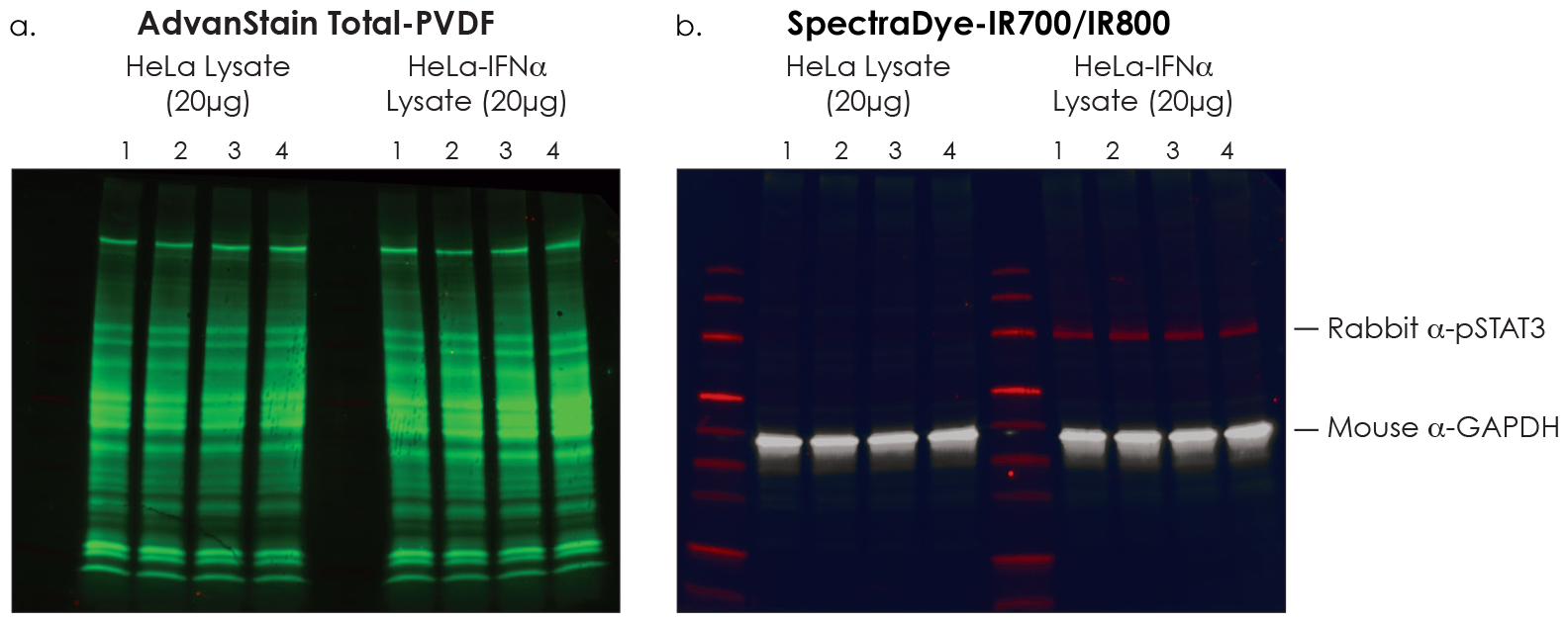
Figure 3. AdvanStain Total is compatible with IR fluorescent Western blotting, without destaining. Untreated and IFNα treated HeLa lysate was loaded in quadruplicate then transferred to a PVDF membrane using a wet transfer. After transfer, the membrane was stained with AdvanStain Total and imaged using a Cy3 filter (a). After staining, the blot was blocked and probed for GAPDH and phosphorylated STAT3. SpectraDye-IR700 and IR800 secondary antibodies were used for detection (b).
Chemiluminescent Western blotting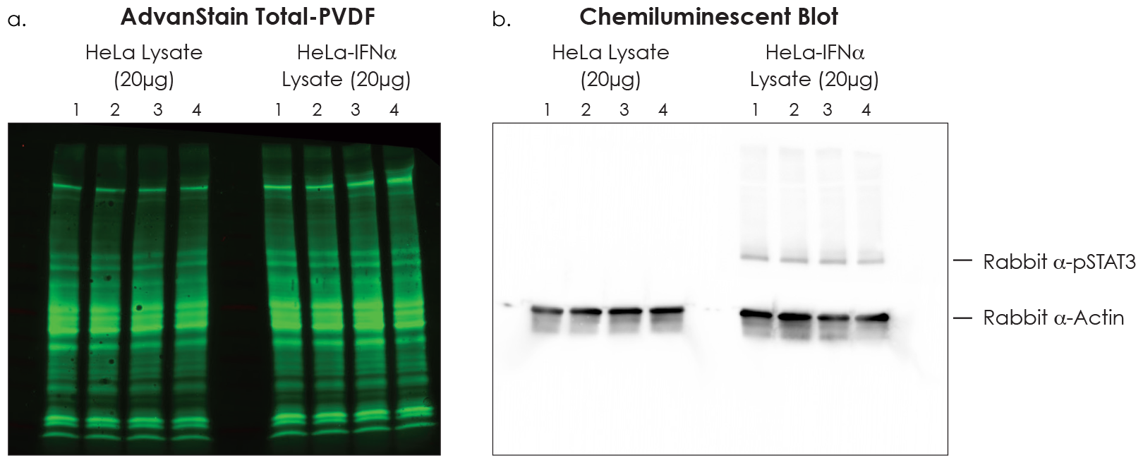
Figure 4. AdvanStain Total is compatible with chemiluminescent Western blotting, without de-staining. Untreated and IFNα treated HeLa lysate was loaded in quadruplicate then transferred to a PVDF membrane using a wet transfer. After transfer, the membrane was stained and imaged using a Cy3 filter (a). After staining the blot was blocked and probed for Actin and phosphorylated STAT3. The antibodies were detected with a goat anti-rabbit HRP conjugate and WesternBright-ECL HRP Substrate (b).
Maximum flexibility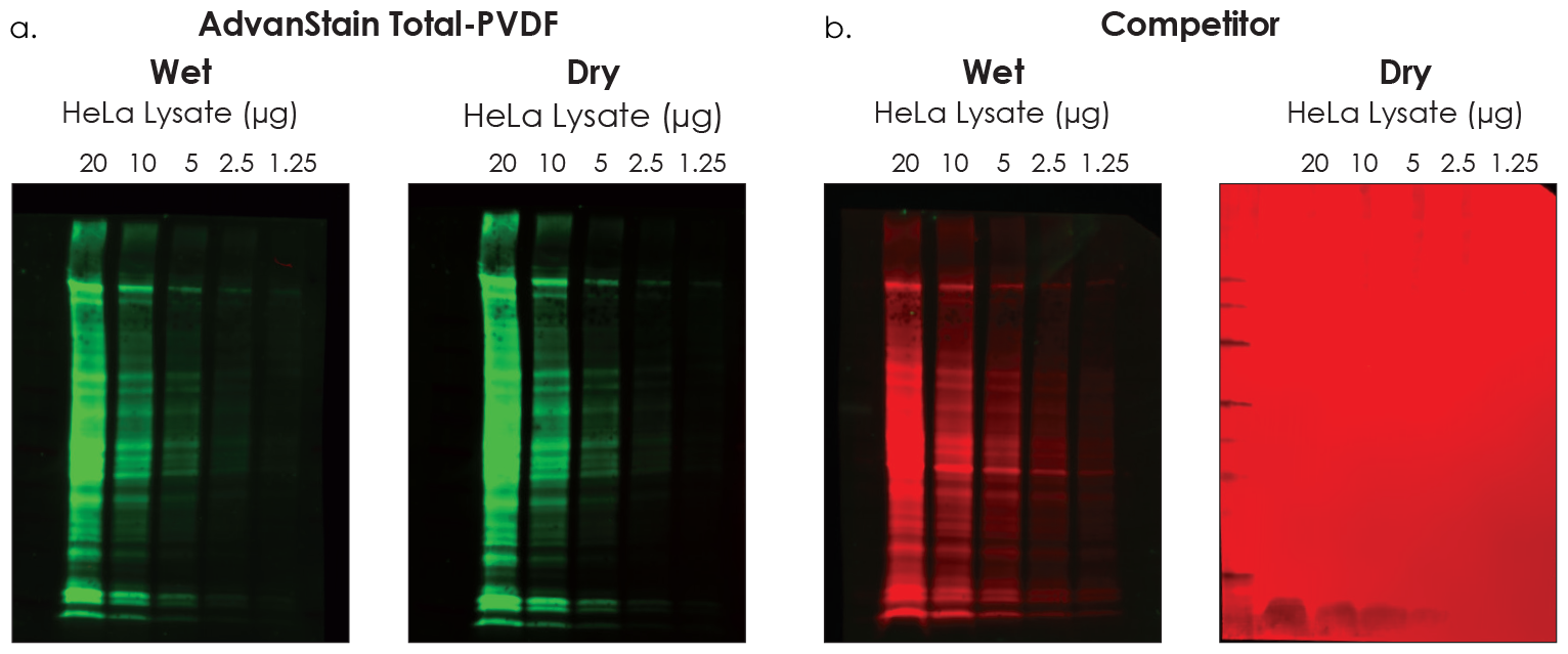
Figure 5. AdvanStain Total may be imaged wet or dry without increasing general background. Dilutions of untreated HeLa lysate were electrophoresed on an SDS-PAGE gel then transferred to a PVDF membrane using a wet transfer. After transfer, the membrane was partitioned and stained. AdvanStain Total was imaged in the Cy3 channel wet and dry (a), the competitor stain was imaged in the Cy5 channel wet and dry (b).
 Click here to request a sample for PVDF membrane
 Click here to request a sample for NC membrane

Connect